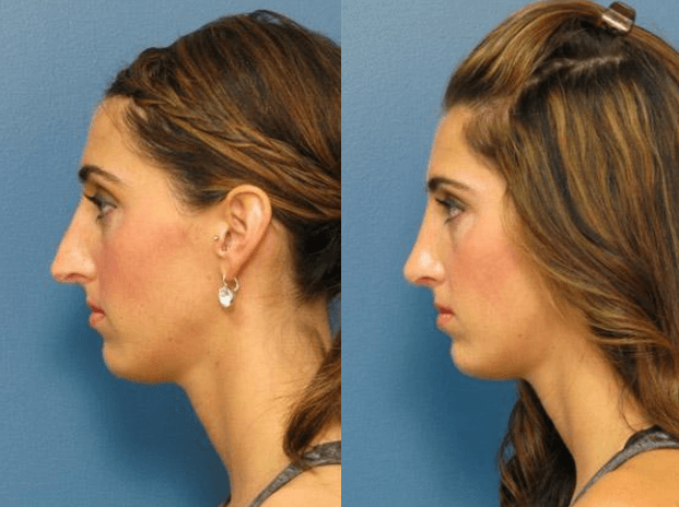
Austin Rhinopasty Surgeon - An Overview
Table of ContentsThe Best Guide To Austin Rhinopasty SurgeonFacts About Rhinoplasty Surgeon Austin RevealedThe 9-Second Trick For Rhinoplasty Surgeon Austin
Based upon the available area of nasal skin, the surgeon chooses the locale for the bilobed flap, and orients the pedicle. If the defect is in the lateral aspect of the nose, the pedicle is based medially. If the problem is at the nasal pointer, or at the nasal dorsum, the pedicle is based laterally.

The outer semi-circle specifies the required length of the two lobes of the skin flap. The inner semi-circle bisects the center of the original injury, and continues across the donor skin, establishing limit measure of the pedicle common to the 2 lobes of the flap. The surgeon then draws 2 lines from the pinnacle of the wound; the very first line drawn is at an angle of 45 degrees from the long axis of the injury, and the 2nd line drawn is at a 90-degree angle from the axis of the wound.
The delineation of each of the 2 lobes of the flap starts and ends at the inner semi-circle, and reaches the outer semi-circle, to the point where it intersects its central axis. The width of the very first lobe is approximately 2 mm narrower than the width of the wound; the width of the second lobe is around 2 mm narrower than the width of the first lobe.
The injury is deepened, to the nasal skeleton, to accommodate the tissue density of the bilobed flap (rhinoplasty austin tx). Technically, cutting the injury, expanding it, is more suitable, and more secure, than cutting (thinning) the flap to fit the injury. Undermining the donor site for the 2nd lobe permits closing it mostly; it likewise eliminates excess-skin "dog-ears" at the donor site.
II. Nasolabial flap In the 19th century, the surgical methods of J.F. Dieffenbach (17921847) promoted the nasolabial flap for nasal reconstruction, for which it stays a fundamental nose surgical treatment procedure. The nasolabial flap can be either superiorly based or inferiorly based; of which the superiorly based flap is the more useful rhinoplastic application, since it has a more versatile arc of rotation, and the donor-site scar is unnoticeable.
The Main Principles Of Rhinoplasty Surgeon Austin
The blood supply for the flap pedicle are the transverse branches of the contralateral angular artery (the facial artery terminus parallel to the nose), and by a confluence of capillary from the angular artery and from the supraorbital artery in the median canthus, (the angles formed by the meeting of the upper and lower eyelids) (rhinoplasty austin tx).
The nasolabial flap is a random flap that is emplaced with the proximal (near) part resting upon the lateral wall of the nose, and the distal (far) part resting upon the cheek, which includes the main angular artery, therefore is perfused with retrograde arterial circulation. Surgical strategy the nasolabial flap The pedicle of the nasolabial flap rests upon the lateral nasal wall, and is shifted a maximum of 60 degrees, in order to avoid the "bridge impact" of article a flap emplaced across the nasofacial angle.
The shape of the skin flap is cut from the wound template fabricated by the cosmetic surgeon. An incision is made to the flap (without an anaesthetic injection of epinephrine), which then is elevated and oriented, in an inferior-to-superior instructions, between the subcutaneous fat and the muscle fascia. The cutting continues until the skin flap can be freely transposed upon the nasal flaw.
The flap then is bent back (shown), and can be thinned (cut) under loupe zoom; however, a nasolabial flap can not be thinned as quickly as an axial skin-flap. rhinoplasty surgeon austin. After the nasolabial flap has been emplaced, the flap donor-site injury is sutured closed. For a wound of the lateral nasal wall that is less than 15 mm broad, the flap donor-site can be closed mainly, with sutures.
Such threats are avoided by advancing (moving) the skin of the cheek towards the nasofacial junction, where it is sutured to the deep tissues. In addition, a narrow injury, less than 1 mm wide can be permitted to recover by secondary intent (self-governing re-epithelialisation). III. The paramedian forehead flap The paramedian forehead flap is the premier autologous skin graft for the restoration of a nose, by replacing any of the aesthetic nasal subunits, specifically relating to the problems of different tissue thickness and click over here now skin color.
Limited length is an useful application limit of the paramedian forehead flap, particularly when the client has a low frontal hairline. In such a client, a little portion of scalp skin can be included to the flap, however it does have a various skin texture and does continue growing hair; such mismatching is prevented with the transverse emplacement of the flap along the hairline; yet that part of the skin flap is random, therefore risks a greater incidence of necrosis.
Unknown Facts About Rhinoplasty Austin Tx
Nevertheless, the second phase of the nasal restoration can be performed with the patient under local anaesthesia. Visually, although the flap donor-site scar heals well, it is visible, and thus tough to hide, particularly in men. Nose surgery: A paramedian forehead flap design. Surgical strategy the paramedian forehead flap The surgeon creates the paramedian forehead flap from a custom-fabricated three-dimensional metal foil template originated from the measures of the nasal flaw to be corrected.
Later on, the distal half of the find flap is dissected and thinned to the subdermal plexus. The surgeon produces a metal foil template stemmed from the measurements of the nasal injury. Applying a Doppler ultrasonic scanner, the surgeon identifies the axial pedicle of the tissue-flap (made up of the supraorbital artery and the supratrochlear artery), typically at the base, next to the median eyebrow; the point typically is between the midline and the supraorbital notch.